
The world’s first crystal-based detector, a perovskite camera, can see inside the human body.
This new research will soon be able to change the way doctors can see inside the human body.
According to SciTechDaily, it was developed by scientists in both America and China. This new device is able to capture gamma rays with unprecedented clarity, so it promises faster, safer, and more affordable scans.
This new detector’s goal is to reduce costs while also improving the quality of nuclear medicine.
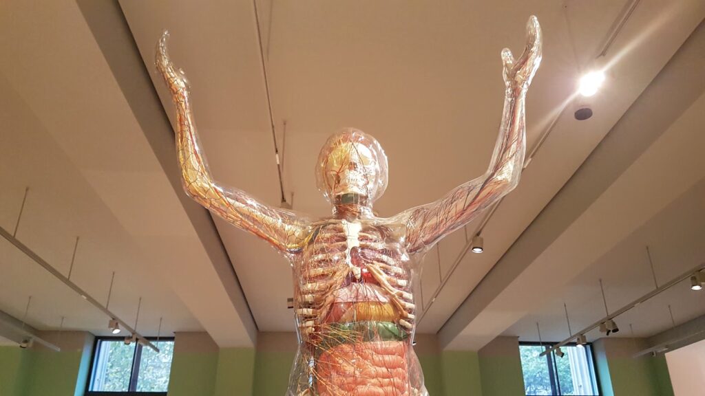
These days, doctors use nuclear medical techniques to observe how the heart pumps, as well as to follow patterns of blood flow and to identify diseases that are hidden deep within the human body.
Currect detectors are falling short
Scanners that are already in place rely on detectors that are costly and difficult to make and manufacture. As a result, current detectors tend to fall short.
Researchers from Northwestern University and from Soochow University (in China) have found a solution to this with the first pervoskite-based detector, which is able to capture gamma rays with exact precision for imaging.
Benefits of this new technological innovation
This innovation has the ability to make nuclear imaging methods more accurate, efficient, affordable, and safe.
From a patient standpoint, the advantages can be shorter scanning sessions, clearer diagnostic images, and lower exposure to radiation, which seems like a win-win-win for all involved.
This study was initially published in the scientific journal “Nature Communications.”
Mercouri Kanatzidis is a Professor of Chemistry at Northwestern University, and he indicated that “perovskites are a family of cystals” that are known for “transforming the field of energy” and now, they can do the same for “nuclear medicine.”
“This is the first clear proof that perovskite detectors can produce the kind of sharp, reliable images that doctors need to provide the best care for their patients,” Kanatzidis said.
Turning to perovskite crystals
To address these challenges with current detectors, the research team turned to perovskite crystals, which Kanatzidis has been studying for over 10 years.
In 2012, Kanatzidis and his group created the first solid-film solar cells using perovskites. The following year, in 2013, he showed that single perovskite crystals could effectively detect X-rays and gamma rays.
“When we first discovered in 2013 that perovskite single crystals could detect X-rays and gamma rays, we could only imagine their potential,” Kanatzidis recalled.
This advance set off a wave of international research and helped establish a new field focused on radiation detection materials.
“Now, we’re showing that perovskite-based detectors can deliver the resolution and sensitivity needed for demanding applications like nuclear medicine imaging. It’s exciting to see this technology moving closer to real-world impact,” Kanatzidis elaborated.
By carefully growing and shaping these crystals, the researchers created a pixelated sensor — just like the pixels in a smartphone camera — that delivers record-breaking clarity and stability.
This way, they can pave the way for practical integration into next-generation nuclear medicine imaging systems.
Real-world impact and commercialization
The Northwestern spinout company Actinia Inc. is commercializing this new technology, where it is working with partners in the medical device field to bring it out of the lab and into hospitals.
“High-quality nuclear medicine shouldn’t be limited to hospitals that can afford the most expensive equipment,” Kanatzidis acknowledged.
“With perovskites, we can open the door to clearer, faster, safer scans for many more patients around the world. The ultimate goal is better scans, better diagnoses, and better care for patients,” he elaborated.
Jamie Prevatt, RN, who has been a nurse in the United States for 33 years, remarked, “This new medical technology innovation sounds amazing. This advancement will help with diagnosing, is less evasive, and it will certainly help with patient recovery.”